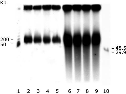
Figure 6.
Characterization of DNA heterogeneity in mitochondrial nucleoids by PFGE. Three agarose-imbedded nucleoids samples, each isolated from 2 mg protein equivalent of mitochondria, were incubated with self-digested proteinase K at a concentration of 0.2, 1 and 5 mg/ml and loaded in lanes 2, 3 and 4, respectively. An identical nucleoids sample was incubated with 1 mg/ml of proteinase K (without self-digestion) and loaded in lane 5. Three agarose-imbedded mitochondrial samples (2 mg protein equivalent each) were incubated with self-digested proteinase K at a concentration of 0.2, 1 and 5 mg/ml and loaded in lanes 6, 7 and 8, respectively. Another identical mitochondrial sample was incubated with 1 mg/ml proteinase K (without self-digestion) and loaded in lane 9. After PFGE, the gel was blotted and hybridized with a 32P-labeled probe of pure mtDNA. Exposure time was 10 h for lanes 2–5 and 5 h for lanes 6–9. Size markers are in lanes 1 and 10. The same result was obtained with a 32P-labeled probe obtained from a PCR-amplified cox 3-gene fragment (data not shown).

