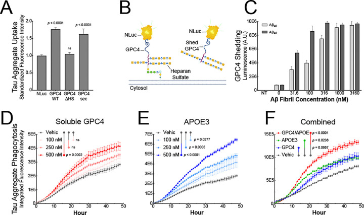
Figure 6: β-Amyloid fibrils lead to GPC4 shedding and APOE secretion which promote tau phagocytosis.
(A) GPC4 WT, GPC4-ΔHS, GPC4-sec, or NLuc control plasmids were transiently transfected into HEK293T cells and tau aggregate-AF647 uptake was measured via flow cytometry. (B) Schematic model depicting NLuc-GPC4 fusion protein for tracking GPC4 shedding into the conditioned media via luminescence. (C) NLuc-GPC4 luminescence was measured in iTF Microglia conditioned media after a 24 h treatment with Aβ40 and Aβ42 fibrils. pHrodo-red labeled tau aggregates (50 nM) were preincubated with (D) soluble recombinant GPC4 or (E) soluble recombinant APOE3 and uptake was measured every hour for 48 h with an Incucyte SX5. (F) pHrodo-red labeled tau aggregates (50 nM) were preincubated with soluble recombinant GPC4 alone, APOE3 alone, or GPC4 + APOE3 (250 nM) and uptake was measured every hour for 48 h. The statistical analyses were performed with a one-way ANOVA and Holm-Sidak multiple comparisons test. N = 4. The data represent the means ± SEM.

