
FIGURE 4.
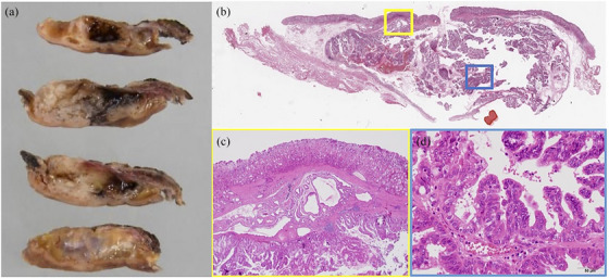
The resected specimen of the gastric lesion. (a) The tumor was 30 × 23 mm in size. Macroscopic findings showed cystic lesions with hemorrhage and mucus. (b) Hematoxylin‐stained loupe image showed that the tumor exhibited an inverted growth pattern with compression of the submucosa. (c) Heterotopic submucosal gastric glands (HSG) were observed around the tumor. (d) The dilated heterotopic submucosal gastric glands manifested a cyst‐like component, in which papillary proliferation of atypical cells exhibiting spindle‐shaped to round nuclei was evident.