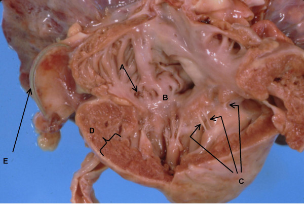
Figure 1.

Anatomy of right ventricle and atrium. A. Dilated coronary sinus. B. Dysplastic tricuspid valve. C. Short thickened chordae tendinae almost implanted into papillary muscle. D. Right ventricular hypertrophy with ventricular wall thickness of 8 mm. E. Pacer wire.
