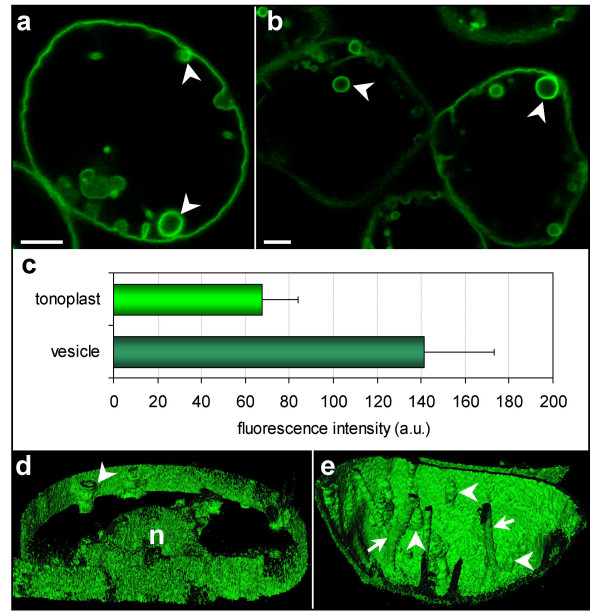
Figure 6.
PEG-stressed cells. (a, b) Localization of BobTIP26-1::GFP in the membrane of spherical structures (arrowheads) within vacuoles. Bar = 10 μm. (c) A histogram of fluorescent intensity values collected from both the tonoplast of the cell periphery and vesicles. (d) A 3-D slice reconstruction through the vacuole at the position of the nucleus (n). Arrowhead = spherical structure. (e) 3-D reconstruction through a part of a vacuole. Transvacuolar strands are still present in these cells (arrow); spherical structures are primarily fixed onto the tonoplast (arrowheads).

