
Figure 3.
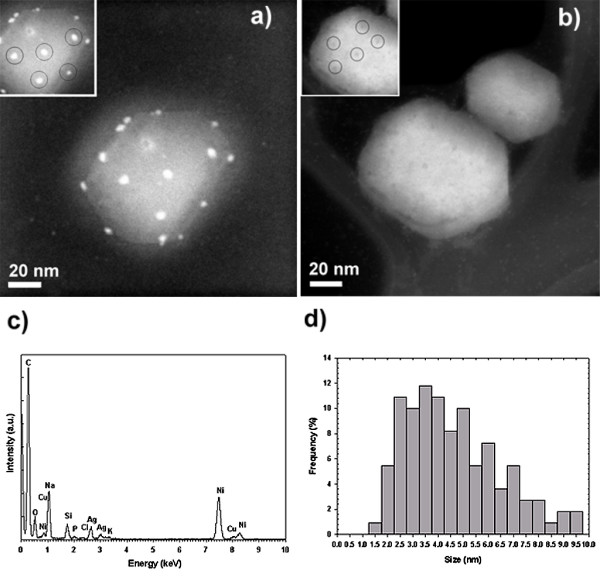
HAADF images of the HIV-1 virus. a) HAADF image of an HIV-1 virus exposed to BSA-conjugated silver nanoparticles. Inset shows the regular spatial arrangement between groups of three nanoparticles. b) HAADF image of HIV-1 viruses without silver nanoparticle treatment. Inset highlight the regular spatial arrangement observed on the surface of the untreated HIV-1 virus. c) EDS analysis of image a) confirming the presence of Ag. The C signal comes from both the TEM grid and the virus, O, and P are from the virus, and Na, Cl, and K are present in the culture medium. Ni and Si come from the TEM grid, while Cu is attributed to the sample holder. d) Composite size distribution of silver nanoparticles bound to the HIV-1 virus, derived from all tested preparations.