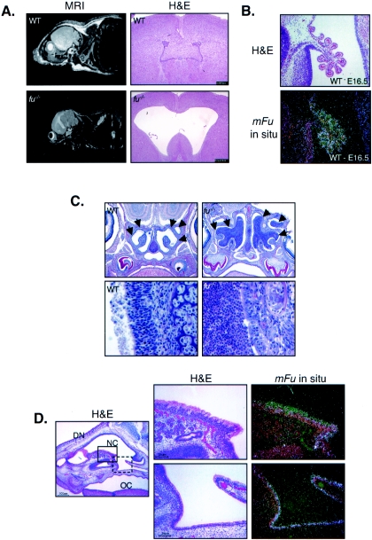
FIG. 3.
Areas of high fused expression correlate with hydrocephalus and suppurative rhinitis. (A) fused knockout mice develop a communicating form of hydrocephalus. Wild-type (WT) and fused knockout (fu−/−) mice are shown by MRI on the left, and brain sections stained with H&E are shown on the right. fu−/− mice develop a progressive, communicating form of hydrocephalus. (B) The fused mRNA is expressed highly in the choroid plexus. The top image shows H&E staining of an E16.5 mouse brain, while the bottom image shows the anti-fused radiolabeled in situexposure (white areas are positively stained with fused antisense probes). (C) fu−/− mice develop bilateral suppurative rhinitis characterized by massive infiltration by neutrophils. The top images show saggital sections of WT and fu−/− nasal cavities at a magnification of ×10, whereas the lower images show the nasal epithelial border in WT and fu−/− mice at a magnification of ×40. WT nasal cavities have a well-defined border at the nasal epithelium with no infiltrating cells (arrows), whereas fu−/− nasal cavities are filled with infiltrating neutrophils (arrows). (D) The fused mRNA is expressed in normal nasal epithelium. The leftmost image is an H&E-stained section at a magnification of ×10. The dorsum of the nose (DN), the nasal cavity (NC), and the oral cavity (OC) are indicated. The solid box corresponds to the H&E and fused radioisotopic in situ hybridization images (magnification, ×40) in the top right images, whereas the dashed box corresponds to the bottom right images.

