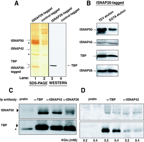
FIG. 4.
TBP is associated with tSNAPc. (A) A silver-stained SDS-10% polyacrylamide gel of affinity-purified proteins from a TAP-tagged tSNAP26 (lane 1) or a control TAP-tagged (lane 2) strain. Nonspecific proteins observed in both lanes are marked by asterisks. Lanes 3 and 4 show a Western blot of the corresponding proteins, probed with anti-TBP antibody. (B) Western analysis of proteins from TAP-tSNAP26 extracts after sequential IgG (lane 1) and calmodulin affinity (lane 2) purification, probed with the indicated antibodies. (C) Western analysis of tSNAP50 and TBP after immunoprecipitation (Ip) from wild-type T. brucei extracts by antisera (α-) as indicated above each lane. The asterisk indicates antibody light chain. (D) Western analysis of tSNAP50 and TBP after immunoprecipitation from wild-type T. brucei extracts with the antisera at different salt concentrations. Total amounts of precipitated protein are less at 0.4 M salt than at 0.2 M, probably reflecting reduced antibody-antigen affinity. PreIm, preimmune.

