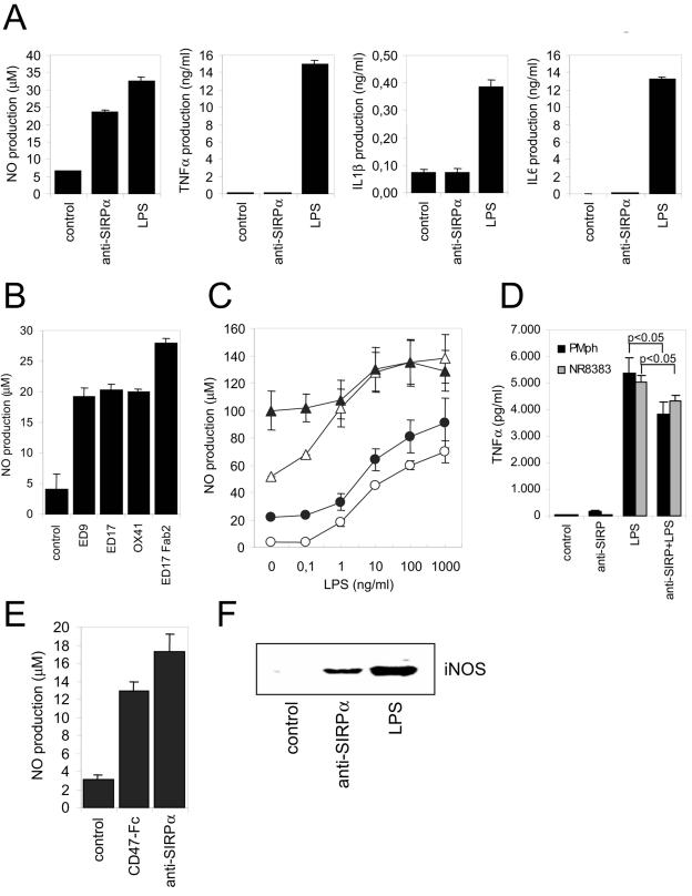
FIG. 1.
Ligation of SIRPα induces macrophage NO production. (A) Rat NR8383 macrophages were cultured for 18 h. in the presence of control IgG1 (MAb BF5, 20 μg/ml), anti-SIRPα MAb ED9 (20 μg/ml), or LPS (100 ng/ml), and amounts of secreted NO, TNF-α, IL-1β, or IL-6 were determined. (B) NO production in NR8383 macrophages was measured after addition of different intact MAb or corresponding F(ab)2 fragments (all at 20 μg/ml; 18 h of incubation). (C) Effects of control IgG1 (20 μg/ml, ○), anti-SIRPα MAb ED9 (20 μg/ml, •), IFN-γ (20 U/ml, ▵), or ED9 plus IFN-γ (▴) either alone, or in combination with various LPS concentrations, on NO production in rat peritoneal macrophages. (D) TNF-α secretion by peritoneal macrophages (PMph) or NR8383 cells cultured in the presence or absence of ED9 (20 μg/ml) and/or LPS (100 ng/ml) for 18 h (P values were obtained by Student's t test). (E) NO production in NR8383 macrophages triggered by murine CD47-Fc protein (25 μg/ml; 18 h of incubation) or anti-SIRPα MAb ED9 (10 μg/ml). (F) Anti-SIRPα MAb ED9 (10 μg/ml; 18 h of incubation) induces iNOS protein expression in NR8383 macrophages as shown by Western blotting. Experiments were performed at least in triplicate, and results are shown as means ± SD.

