FIG. 6.
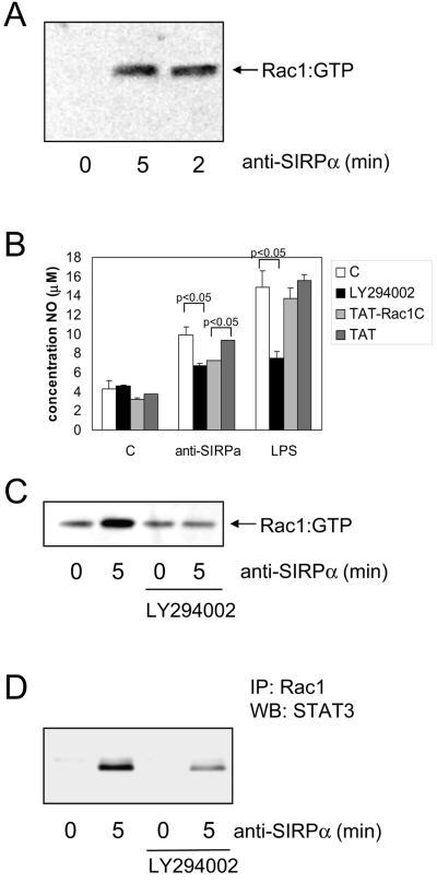
Role of Rac1 and PI3-K in SIRPα-induced NO production in macrophages. (A) Rac1 activation was determined (see Materials and Methods for details) in NR8383 cells following SIRPα ligation using MAb ED9 (10 μg/ml). (B) NR8383 cells in the presence of LY294002 (40 μM; 30-min preincubation), cell permeable TAT-Rac1 C-terminal peptides (200 μM; 5-min preincubation), or control TAT peptide (200 μM) were subsequently stimulated by anti-SIRPα (10 μg/ml) or LPS (100 ng/ml). After 20 h, NO production in the supernatants was measured. Shown are results from a representative experiment performed in triplicate (means ± SD). The differences between control and LY294002 in the anti-SIRPα- and LPS-treated samples and between Tat and Tat-Rac1 in the anti-SIRPα-treated samples are statistically significant (P < 0.05 by Student's t test). (C and D) NR8383 cells were pretreated for 30 min with LY294002 (40 μM) and then stimulated with anti-SIRPα (10 μg/ml) for 5 min and lysed. From these lysates, active Rac1 was pulled down as described in Materials and Methods (C), and subsequently, total Rac1 was immunoprecipitated (IP) and the associated STAT3 shown on a Western blot (WB) (D). Shown are representative results from three experiments.

