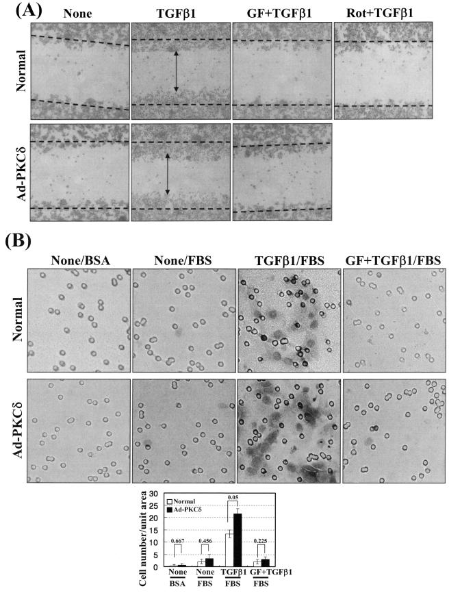
FIG.8.
Wound healing and invasion also depend on TGFβ1, PKCδ, and integrins. (A) TGFβ1- and PKCδ activity-mediated wound healing on Fn. Prior to making wounds, normal or Ad-PKCδ-infected cells were replated onto Fn-precoated six-well culture plates. Twelve hours later, wounds (marked as dotted lines) were created by scraping through the monolayer. Cells were then incubated in the absence or presence of TGFβ1 treatment without or with PKC inhibition (GF-109203X [GF] or rottlerin [Rot]) for 36 h at 37°C, prior to taking images. Note that TGFβ1-mediated healing in PKCδ-overexpressing cells is slightly enhanced, compared to that in normal control cells (i.e., compare healing around the vertical arrowheads). (B) TGFβ1- and PKCδ activity-mediated invasion of the cells through matrigel. Normal or Ad-PKCδ-infected cells in RPMI 1640 containing 1% BSA were replated onto matrigel, prepared as explained in Materials and Methods, and concomitantly treated with or without 5 ng/ml TGFβ1. Certain cells were pretreated with 12.5 μM GF-109203X, 30 min prior to the TGFβ1 treatment. Then the upper chambers were placed into 24-well culture dishes containing 0.6 ml of RPMI 1640 with 10% fetal bovine serum (FBS) or with 1% BSA. After incubation at 37°C for 72 h, invasive cells were fixed and stained with crystal violet, and then images were taken, as explained in Materials and Methods. The images shown are representative of three isolated experiments. Stained cells in three independent images for each condition were counted for the graphic presentation (mean ± standard deviation).

