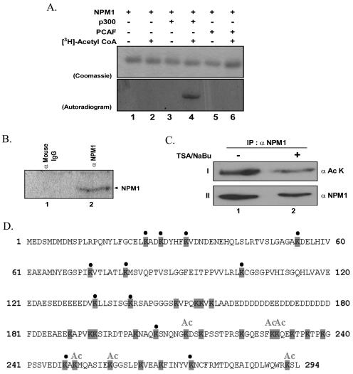
FIG. 4.
Nucleophosmin becomes acetylated both in vitro and in vivo. (A) In vitro gel HAT assay carried out with NPM1 as a substrate without (lanes 1 and 2) or with (lanes 3 and 4) the activity-normalized p300. The acetylation reactions were also tested with PCAF (lanes 5 and 6) as the histone acetyltransferases. Lanes 2, 4, and 6 show the acetylation reaction in the presence and lanes 1, 3, and 5 in the absence of [3H]acetyl-CoA. The gel was then subjected to CBB staining and fluorography and exposed to an X-ray film. Top panel shows the CBB-stained gel, while the bottom panel shows the autoradiogram. (B) In vivo acetylation of nucleophosmin. Nucleophosmin was immunoprecipitated with the monoclonal antibody (lane 2) from 293T cells treated with deacetylase inhibitors (500 ng/ml TSA and 5 mM sodium butyrate) as well as [3H]sodium acetate. The immunoprecipitate was analyzed on an SDS-12% PAGE gel and processed as described for panel A. Mouse preimmune serum was used as an immunoprecipitation control (lane 1). IgG, immunoglobulin G. (C) In vivo acetylation of nucleophosmin under normal growth conditions. Nucleophosmin was immunoprecipitated (IP) with the monoclonal antibody from 293T cells treated with deacetylase inhibitors (500 ng/ml TSA and 5 mM sodium butyrate). The immunoprecipitate was analyzed on an SDS-12% PAGE gel, blotted, and probed with antibody against acetylated lysine (panel I). The lysates used in the pulldown assay were analyzed on an SDS-12% PAGE gel, blotted, and probed with the antibody against NPM1 (panel II). (D) Protein sequence of nucleophosmin showing the acetylation sites. The lysine residues are shaded. The lysines that are acetylated are marked “Ac,” while the ones that are not acetylated are marked with a bullet. Lysines that could not be assigned as either of the above have been left unmarked.

