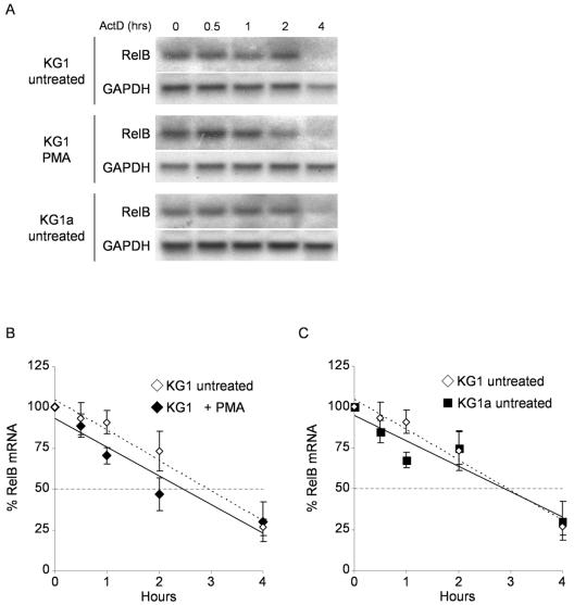
FIG. 3.
RelB expression is not regulated by changes in mRNA stability. (A) Determination of RelB mRNA half-life in KG1, KG1a, and PMA-treated KG1 cells. KG1 and KG1a cells left unstimulated or stimulated with PMA for 2 h were treated with actinomycin D to arrest transcription before isolation of total RNA at the indicated time points. RelB and GAPDH transcripts were detected in Northern blotting assays as previously described. (B) PMA treatment does not increase the RelB mRNA half-life in KG1 cells. Hybridization signals for the RelB and GAPDH mRNAs were quantified by phosphorimaging in KG1 cells left untreated or treated with PMA for 2 h. Each lane was normalized for equal loading by dividing the RelB content by the respective GAPDH signal and expressed relative to RelB content at 0 h for each condition. The mRNA half-life was calculated by linear regression analysis to be 3.0 h and 2.5 h for untreated and PMA-treated KG1 cells, respectively. The dashed line parallel to the x axis represents 50% mRNA decay. The data represent the mean ± the standard error from two independent experiments. (C) RelB mRNA half-life is the same in KG1 and KG1a untreated cells. A RelB mRNA half-life of 3.0 h was determined for untreated KG1a cells as described for panel B. The data represent the mean ± the standard error from two independent experiments.

