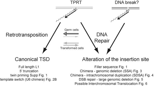
FIG. 8.
Competing pathways for L1 retrotransposition in cultured human cells. L1 retrotransposition can be initiated in two ways. The major pathway uses L1 EN to initiate TPRT (left panel). A secondary pathway could use a DNA lesion to initiate reverse transcription (right panel) (56). We propose that the resultant cDNA intermediate can be resolved using distinct DNA repair pathways (see Discussion). “Canonical” retrotransposition (left panel) results in the formation of L1 insertions with standard hallmarks of TPRT. “Abortive” retrotransposition (right panel) can lead to a variety of structures, depending on the DNA repair pathway that is utilized to resolve the L1 cDNA intermediates. The balance between “conventional” and “abortive” retrotransposition likely depends on the cellular milieu. Conventional retrotransposition may be more apparent in germ cells (depicted by a large horizontal gray arrow over a thin light gray arrow), whereas “abortive” retrotransposition may be more apparent in transformed cultured cells (depicted with equally sized gray horizontal arrows).

