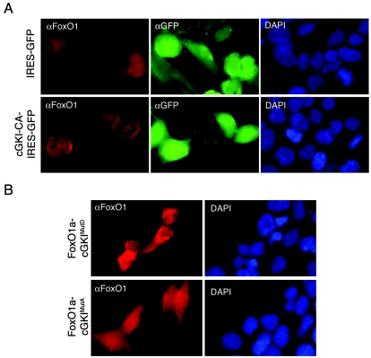
FIG. 8.
cGKI phosphorylation provokes exclusion of FoxO1a from the nuclei of 293T cells. (A) Immunofluorescence analyses of 293T cells stably expressing cGKI-CA and then transduced with a FoxO1a-expressing retrovirus show cytoplasmic accumulation of FoxO1A. GFP fluorescence demarks transduced cells. 4′,6′-Diamidino-2-phenylindole (DAPI) staining reveals cell nuclei. (B) Localization of FoxO1a in 293T cells expressing FoxO1a mutated at the cGKI phosphorylation sites. FoxO1a-cGKIMutD-expressing (phosphomimetic) cells show predominantly cytoplasmic staining, while FoxO1a-cGKIMutA (nonphosphorylatable) expression is diffuse, similar to that in cells overexpressing wild-type FoxO1a. DAPI staining again demarks all nuclei in the field.

