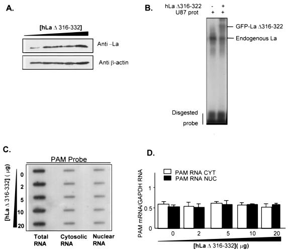
FIG. 8.
Effect of high expression of hLaΔ316-332 on subcellular localization of PAM mRNA. U87 cells were transfected with increasing amounts of the hLaΔ316-332 plasmid (0, 2, 5, 10, and 20 μg), and 24 or 48 h later, cells were homogenized to prepare RNA and protein extracts, respectively. (A) hLaΔ316-332 protein levels in total extract were detected by Western blotting. β-Actin levels were monitored as controls for loading. (B) Representative gel mobility shift assay with U87 cytoplasmic extracts transfected with 20 μg of either empty vector (lane 1) or GFP-hLaΔ316-332 plasmid (lane 2). (C) PAM mRNA levels in total, cytosolic, and nuclear RNA were analyzed by slot blot and quantified as described in the legend of Fig. 7. (D) Levels of PAM mRNA were normalized to GAPDH mRNA levels. (E) Levels of nuclear PAM RNA detected with intronic PAM probe were normalized to levels of nuclear U6 RNA. CYT, cytosolic RNA; NUC, nuclear RNA.


