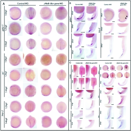
FIG. 4.
Whole-mount in situ analysis of various hematopoietic and vascular markers. Expression of various hematopoietic genes in control and in zMelk-like gene MO-injected embryos. (A) Whole-mount in situ hybridization of gata-1, scl, and lmo-2. Lateral view with anterior at the top (first and third columns from the left) and dorsal view with anterior at the top (second and fourth column from the left) of probe-hybridized zebra fish embryos injected with either control MO or zMelk-like gene MO are shown. Scale bars, 100 μm. (B) Lateral views of whole-mount control MO- or zMelk-like gene MO-injected embryos at 16 to 26 hpf in situ hybridized for gata-1, scl, and lmo-2 mRNA. Scale bars, 150 μm. (C) Embryos injected with control MO or zMelk-like gene MO were harvested at the indicated stages, and whole-mount in situ hybridization was done by using hoxb6b, c-myb, tie-1, cdx-4, fli-1, and flk-1 probes. Expression patterns of hoxb6b at 19 and 24 hpf (lateral view with dorsal to right and dorsal view with anterior at the top), c-myb at 19 and 24 hpf (lateral view of ICM region), tie-1 at 20 and 24 hpf (lateral view of ICM region), cdx-4 at 12 hpf, 14 hpf (lateral view with anterior to left and posterior view with dorsal at the top), cdx-4 at 19 hpf (lateral view of ICM region), fli-1 at 20 and 24 hpf (lateral views of ICM region) and flk-1 at 24 and 28 hpf (lateral view of ICM region) are shown. Results of antisense probe hybridization are shown in the figure. Sense probe control experiments were done for all of the probes, and no significant signals were detected. Scale bars, 100 μm.

