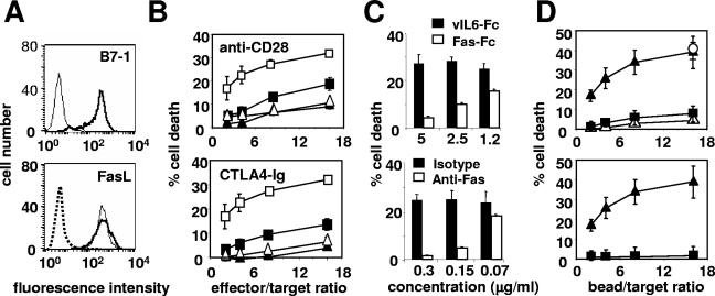
FIG. 1.
Fas-mediated cell death is amplified upon CD28 costimulation. (A) The membrane expression of B7-1 (upper panel) and of FasL (lower panel) on the 1A12 cell line (thin line) and on the 1A12-B7-1 cell line (thick line) was analyzed by flow cytometry. The dotted line depicts an isotype control MAb. (B) 1A12 (▴ and ▵) or 1A12-B7-1 (▪ and □) cell lines were incubated with 51Cr-labeled Jurkat 77 target cells at the indicated ratios in the presence of (▴ and ▪) soluble blocking anti-CD28 MAb (upper panel) or CTLA4-Ig (lower panel) or of (▵ and □) the negative control MAb or the control vIL6-Fc. (C) The 1A12-B7-1 cell line was incubated with the 51Cr-labeled Jurkat 77 target cells at an 8:1 ratio in the presence of Fas-Fc or vIL6-Fc (upper panel) or of the blocking anti-Fas MAb ZB4 or the control MAb (lower panel) at the indicated concentrations. (D) Polystyrene beads were coated with antibodies and mixed for 4 h with 51Cr-labeled Jurkat 77 target cells at the indicated ratios before cell death measurement. In the upper panel, anti-Fas MAb 7C11 (0.08 μg/ml) was coated together with the negative IgG control MAb (▪), the anti-CD28 MAb (▴), or the anti-CD71 MAb (▵) at 10 μg/ml. To quantify the maximum value of Fas-mediated apoptosis in this assay, beads were coated with 7C11 at high concentration (10 μg/ml) (○). In the lower panel, beads were coated with either the anti-Fas MAb 7C11 (0.08 μg/ml) and the anti-CD28 MAb (▴) or the negative IgM control MAb (0.08 μg/ml) and the anti-CD28 MAb (▪). Results represent the means and errors of three independently repeated experiments.

