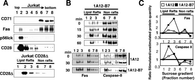
FIG. 4.
Fas is redistributed in the lipid rafts upon CD28 cosignal. (A) Triton X-100 cell lysates prepared from the Jurkat 77 (upper part) or the Jurkat CD28Δ (lower part) cell lines were separated by ultracentrifugation on a sucrose gradient. Ten micrograms of proteins per lane was loaded, separated by SDS-PAGE, and analyzed by immunoblotting for their content in the indicated molecules. (B) Jurkat cells (50 × 106) were incubated at the indicated time with the 1A12 or 1A12-B7-1 cell lines as indicated (ratio, 1:2) and lysed. The lipid rafts were isolated by ultracentrifugation on a sucrose gradient, and Fas or caspase 8 was analyzed by immunoblotting. (Upper panel) Time-dependent relocation of Fas into the microdomains in response to 1A12-B7-1 cells. (Lower panel) Relocation of Fas and caspase 8 in response to 1A12 or 1A12-B7-1 cells after an incubation of 30 min. (C) Densitometry analysis of Fas and caspase 8 distribution. Values correspond to ratios of the intensity of specific bands at 30 min versus 0 min (see Materials and Methods) with 1A12 (□) or 1A12-B7-1 (▪) cells.

