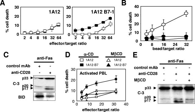
FIG. 9.
CD28 corecruitment increases Fas-mediated apoptosis in primary activated CD4+ T lymphocytes from peripheral blood. (A) 51Cr-labeled activated CD4+ T lymphocytes were incubated with 1A12 or 1A12-B7-1 cells in the absence (▪) or in the presence (□) of the blocking anti-CD28 MAb. (B) 51Cr-labeled activated CD4+ T lymphocytes were incubated with beads coated with the anti-Fas MAb (2 μg/ml) and either the negative IgG control MAb (•) or the anti-CD28 MAb (○). As a control, beads were coated with the anti-CD28 MAb and the negative IgM control MAb (▪). (C) Primary activated CD4+ T lymphocytes were incubated for 2 h with beads coated with the anti-Fas MAb at 2 μg/ml and either the anti-CD28 MAb or an irrelevant IgG MAb. The cells were lysed, and total proteins (10 μg/lane) were separated by SDS-PAGE before immunoblotting for caspase 3 and BID. (D) Primary activated CD4+ T lymphocytes were treated for 20 min with 5 mM MβCD or α-CD and incubated with either 1A12 effector cells or 1A12-B7-1 cells, and a 51Cr release assay was performed. (E) Primary activated CD4+ T lymphocytes were treated for 20 min with 5 mM MβCD or left untreated. After washing, the cells were incubated for 45 min with beads coated with the indicated antibodies and then lysed, and total proteins (10 μg/lane) were separated by SDS-PAGE before immunoblotting for caspase 3. For all of the 51Cr-release assays, results represent the means and errors of three independently repeated experiments.

