FIG. 1.
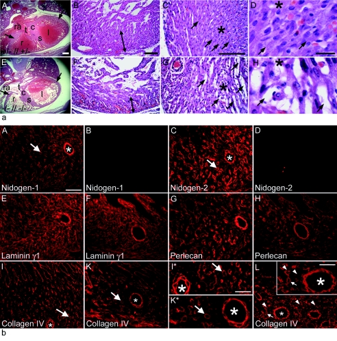
Histology (a) and immunofluorescence microscopy (b) of cardiac tissues from nidogen double-null embryos and neonates. (a) Hematoxylin-and-eosin-stained transverse sections of hearts from control littermates (A to D) and double-null mice (E to H) at E17.5 (A and E) and P0 (B to D and F to H) are shown. The nidogen-deficient hearts (E) were slightly smaller in size than the hearts of control embryos (A). (A, B, E, and F) Arrows, compact zones of the ventricular wall; (C, D, G, and H) asterisks, areas shown at different magnifications; arrows, cell-cell contacts. (A and E) c, endocardial cushion; l, left ventricle; r, right ventricle; ra, right atrial chamber; s, interventricular septum; t, tricuspid valve. Bars, 200 μm (A and E), 50 μm (B, C, F, and G), and 10 μm (D and H). (b) Immunofluorescence was performed on embryonic heart sections at E18.5 using rabbit antisera against nidogen 1 (A and B), nidogen 2 (C and D), laminin γ1 (E and F), perlecan (G and H), and collagen type IV (I, I*, K, K*, and L). Deposition of all basement membrane components is detectable in control sections (A, C, E, G, I, and I*), whereas in mutant cardiac tissues, the staining intensities appeared to be reduced (F, H, K, K*, and L). Areas of the cardiac muscle tissue with smaller (arrows) and larger (asterisks) blood vessels in I and K are shown at a higher magnification in I* and K*. In some sections from nidogen-deficient hearts, collagen IV staining of basement membranes of capillaries (L) appears to vary, with some showing a normal (arrow) and others showing an irregular (arrowheads) or sometimes patchy staining pattern. Bars, 50 μm (A to K, K*, I*, and L) and 25 μm (inset in L).

