
FIGURE 4.
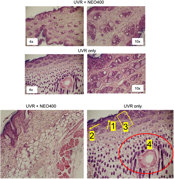
Microscopic appearance of UVR‐exposed skin. Skin sections were prepared from mice subjected to the 5‐week treatment cycle of UVR‐only, or UVR + NEO400 (Schedule A). Histological characteristics were examined under the microscope. As exemplified in these images, the epidermis of UVR‐only skin showed signs of: (1) hypergranulosis, (2) dyskeratosis, (3) hyperplasic epidermis, and (4) abnormal structures with ductal differentiation, which were not apparent in skin sections from mice that had received NEO400 immediately following UVR exposure.