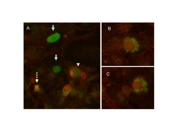
Figure 6.
Immunohistochemical staining of beta-catenin and p53. Cells were treated with 10-11 M 17-beta estradiol for 48 h as described under methods and stained for p53 (green) and beta-catenin (red) using specific antibodies. Panel A shows cells strongly staining for both proteins in the nucleus (broken arrow), strong p53 staining in the nucleus and beta-catenin at the plasma membrane (solid arrows) and strong staining of p53 in the nucleus with beta-catenin relegated to the cytoplasm (arrow head). B and C show apoptotic cells with strong staining of both proteins.

