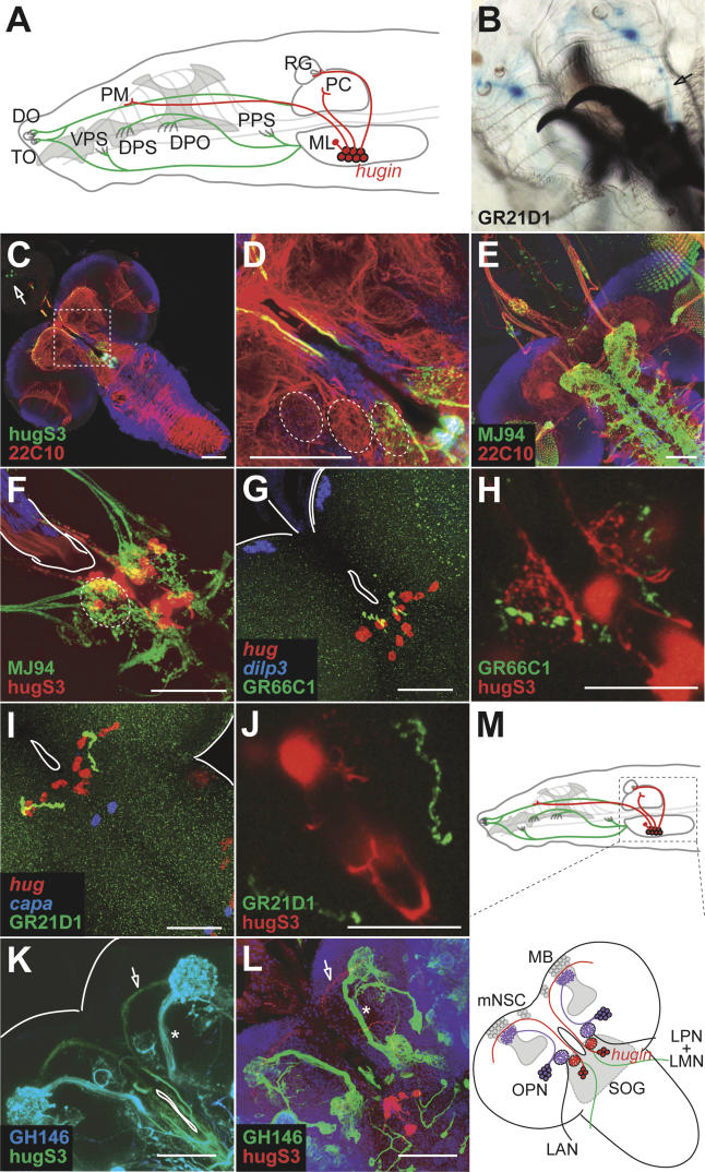
Figure 3. hug Neurons Receive Gustatory Input.
(A) Schematic drawing of the head region of a Drosophila larva with external as well as internal chemosensory neurons in antennomaxillary complex and internal mouth region innervating the larval CNS (depicted in green). hug neurons in the SOG (shown in red) project to the ring gland (RG), pharyngeal muscle (PM) region, and the protocerebrum (PC). Relative positions of external chemosensory sensillae (dorsal organ [do] and terminal organ [to]), internal chemosensory sensillae (ventral pharyngeal sense organ [vps], dorsal pharyngeal sense organ [dps], dorsal pharyngeal organ [dpo], and posterior pharyngeal sense organ [pps]), and major projections to the CNS are shown.
(B) GR21D1-positive sensory neurons (shown by X-Gal staining) in the dorsal organ project axons to the CNS (see arrow).
(C and D) Optical section through median CNS (composed of ten confocal 1-μm sections) shows hug arborizations in the SOG and mushroom body region (shown in green) relative to general neuropil (red) and cortical (DNA marker Draq5, blue) landmarks. Note the labeling of corpora allata cells in the ring gland (arrow; see Materials and Methods and Figure S2). Boxed area is shown at higher magnification in (D), revealing spherically organized neuropil regions lateral to foramen (partially outlined).
(E) Expression of nSyb-GFP under MJ94 enhancer trap construct labels axon terminals of internal gustatory sensory neurons (shown in green), innervating SOG and VNC.
(F) Close-up of SOG region shows co-localization of spherically organized axon terminals of MJ94 positive gustatory neurons (green) and hug neuronal arborizations (red).
(G) Axon terminals of GR66C1-positive chemosensory neurons (shown in green) can be detected in the vicinity of hug cell bodies (shown in red). dilp3 staining (blue) serves as morphological landmark.
(H) Optical section (composed of ten confocal 1-μm sections) containing the GR66C1 axon terminals (shown in green) also include the spherical hug arborizations. hug cell bodies are out of focus in (H); a close-up of the SOG is depicted.
(I and J) Axon terminals of GR21D1 positive chemosensory neurons (shown in green) also project to the vicinity of hug cell bodies (shown in red in [I]), but the optical section comprising these terminals does not contain the hug arborizations (note the absence of spherical hug dendrites in [J] as compared to [H]).
(K–M) Comparison of axon tracts used by olfactory projection neurons, which can be labeled by enhancer trap line GH146 (shown by GFP real color in [K] and in green in [L], marked by asterisks), with those used by hug neurons (shown by YFP real color in [K] and in red in [L], marked by arrows), indicate that hug neurons are distinct from olfactory projection neurons (OPN). These differences are summarized in (M). LAN, larval antennal nerve; LMN, larval maxillary nerve; LPN, larval pharyngeal nerve.

