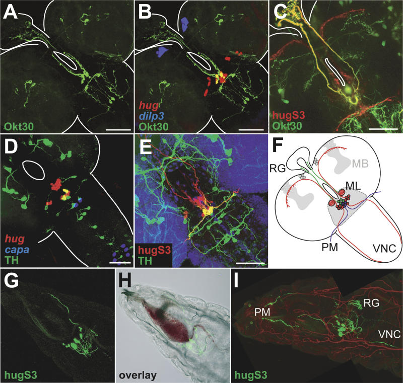
Figure 4. Subpopulations of hug Neurons Innervate Distinct Targets.
(A and B) Enhancer trap line Okt30 (shown in green) labels SOG neurons projecting to ring gland. Okt30 expression pattern colocalizes with hug expression (shown in red in [B]). There are four double positive cells in (B). dilp3 staining (blue) serves as morphological landmark.
(C) Direct detection and false colorization of GFP expressed in Okt30 positive cells (green) and YFP expressed under hug promoter (red) reveals only the axons to ring gland as double positive.
(D) TH promoter construct labels dopaminergic CNS neurons (shown in green). Four TH-positive SOG neurons also express hug (shown in red). capa staining (blue) serves as morphological landmark.
(E) Combination of projection patterns of TH positive cells (green) with hug cells (red) reveals the axons innervating the pharyngeal muscles as the only double positive ones.
(F) Schematic summary of (A–E) showing distinct subpopulations of hug neurons projecting to the ring gland only (green), pharynx only (blue), and the remaining targets (red).
(G–I) Projection pattern of hug is unaffected in klu mutants. Direct detection and false coloring of hug promoter-YFP (shown in green) in the klu background reveals the pharyngeal muscles (PM), the ring gland (RG), and the VNC as being targeted in feeding mutants. Trachea are false-colored (red) in composite figure (I).

