Fig. 4.
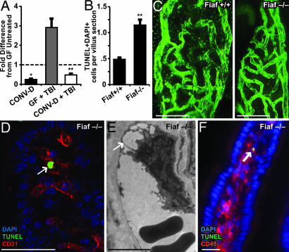
Loss of Fiaf results in loss of resistance to TBI-induced apoptosis in GF mice. (A) Quantitative RT-PCR analysis shows that conventionalization of GF WT mice suppresses Fiaf expression in the distal small intestine and that this effect is not abrogated after 16 Gy of TBI (mean values ± 1 SD are plotted; n = 4 mice per group; *, P < 0.05, compared with GF untreated B6 mice; **, P < 0.01, compared with GF mice treated with 16 Gy of TBI 4 h before they were killed). (B) Quantitative assessment of apoptosis in the villus mesenchyme of GF Fiaf-/- mice and their WT littermates 4 h after 18 Gy of TBI (n = 9 mice per group; ≥100 villus sections assayed per mouse; mean ± 1 SD; **, P < 0.01, compared with GF Fiaf+/+). (C) Single images from digital 3D reconstructions of small intestinal villus capillary networks in GF Fiaf-/- and their WT littermates. (Scale bars, 20 μm.) (D) TUNEL+ CD31+ endothelial cell (arrow) in the small intestinal villus mesenchyme of a GF Fiaf-/- mouse treated as in A. (Scale bar, 50 μm.) (E) TEM image of a villus mesenchymal endothelial cell in a Fiaf-/- mouse killed 4 h after 18 Gy of TBI. Note the nuclear protrusion and avulsion from the basement membrane (arrow), two ultrastructural manifestations of endothelial cell death. (Scale bar, 5 μm.) (F) TUNEL+ CD45+ leukocyte (arrow) in the small intestinal villus mesenchyme of a GF Fiaf-/- mouse treated as in A. (Scale bar, 20 μm.)

