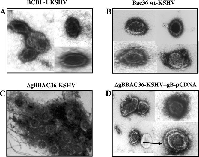
FIG. 10.
Electron micrographs of virus particles. Virus particles in the supernatants of TPA-induced BCBL-1 cells (A), 293T-BAC36 wt cells (B), 293T-ΔgBBAC36 cells (C), and 293T-ΔgBBAC36 cells transfected with gB-pCDNA3.1 plasmid (D) were concentrated, stained with 1% uranyl acetate in distilled water for 30 s, and observed under an electron microscope. Magnifications, ×144,000.

