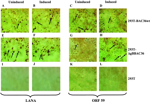
FIG. 4.
Immunoperoxidase assay detecting KSHV ORF 73 (LANA) and ORF59 proteins. 293T, 293T-BAC36wt, and 293T-ΔgBBAC36 cells in chamber slides were uninduced (A, C, E, G, I, and K) or induced with 20 ng/ml of TPA for 96 h (B, D, F, H, J, and L), fixed with ice-cold acetone, and treated with 5% hydrogen peroxide to remove endogenous peroxidase. The cells were incubated at room temperature with rabbit anti-ORF 73 antibodies (A, B, E, F, I, and J) or with anti-ORF59 monoclonal antibodies (C, D, G, H, K, and L), washed, and treated with biotinylated secondary antibody followed by ABC reagent. Color reaction was performed with hydrogen peroxide and diaminobenzidine. The arrows indicate the cells stained with the respective antibodies. The total number of cells and the number of cells stained positive by the reaction were counted in at least three separate areas, and the percentage of cells expressing the respective proteins was calculated. Magnification, ×40.

