FIG. 9.
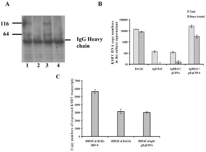
Complementation of ΔgBBAC with gB. (A) Monolayers of 293T-ΔgBBAC36 cells were transfected with gB-pCDNA3.1 plasmid or pCDNA3.1 plasmid for 48 h and induced with TPA for 96 h, and lysates were prepared in RIPA lysis buffer. KSHV gB was immunoprecipitated with rabbit anti-gB polyclonal antibodies, Western blotted, reacted with anti-gB antibodies, and tested with alkaline phosphatase-conjugated secondary antibodies (lanes 3 and 4). Lysates from untransfected 293T-BAC36wt and 293T-ΔgBBAC36 cells induced with TPA for 96 h were also immunoprecipitated and Western blotted (lanes 1 and 2). The numbers indicate the molecular masses (in kilodaltons) of the gB proteins detected. (B) Monolayers of 293T-ΔgBBAC36 cells transfected with gB-pCDNA3.1 plasmid or pCDNA3.1 plasmid alone as the control for 48 h and induced with TPA for 96 h. KSHV was concentrated from the culture supernatants, and total DNA was isolated and subjected to real-time DNA PCR to quantitate the ORF 73 copy numbers. Virus stocks were also pretreated with 12 μg of DNase I for 15 min at room temperature. (C) HMVEC-d cells were infected with virus from 293T-ΔgBBAC36 cells complemented with gB-pCDNA3.1 plasmid, and copy numbers of expressed KSHV transcripts were quantitated as in the procedures described in the legend to Fig. 3D. The error bars indicate standard deviations.

