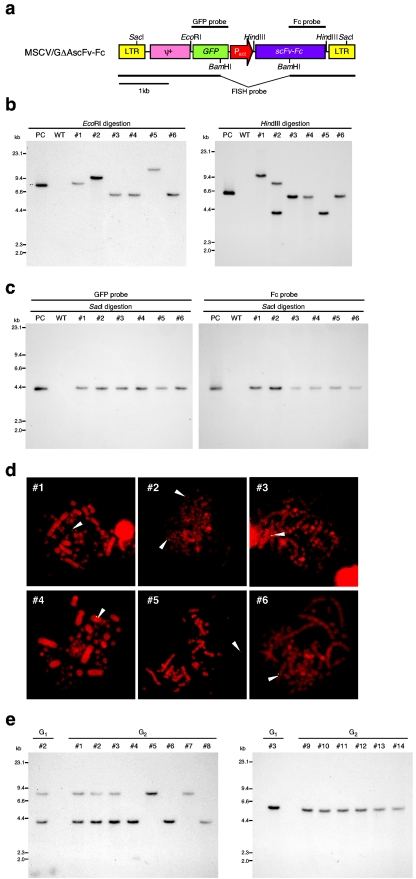
FIG.3.
Analysis of G1 and G2 transgenic chickens expressing scFv-Fc. (a) Restriction enzyme map of the MSCV/GΔAscFv-Fc retroviral vector and locations of probes for Southern and FISH analyses. (b) Determination of the copy number of the transgene in the G1 genome by Southern blot analysis. Genomic DNA extracted from the blood of G1 transgenic chickens was digested with EcoRI (left) or HindIII (right), electrophoresed, and hybridized with the GFP probe. WT, wild type; PC, positive control (pMSCV/GΔAscFv-Fc plasmid). (c) Confirmation of the intactness of the vector sequence in the G1 genome by Southern blot analysis. Genomic DNA fragments digested with SacI were electrophoresed and hybridized with the probe for GFP (left) or Fc (right). (d) FISH analysis for determination of the chromosomal location of the transgene for G1 transgenic chickens. The vector sequence was detected using a fluorescein isothiocyanate-labeled probe prepared from the pMSCV/GΔAscFv-Fc plasmid by digestion with BamHI. The arrowheads indicate the location of the transgene. (e) Southern blot analysis for G2 transgenic chickens derived from G1 transgenic chickens 2 (left) and 3 (right). Genomic DNA extracted from the blood of G2 transgenic chickens was digested with HindIII, electrophoresed, and hybridized with the GFP probe.

