FIG. 3.
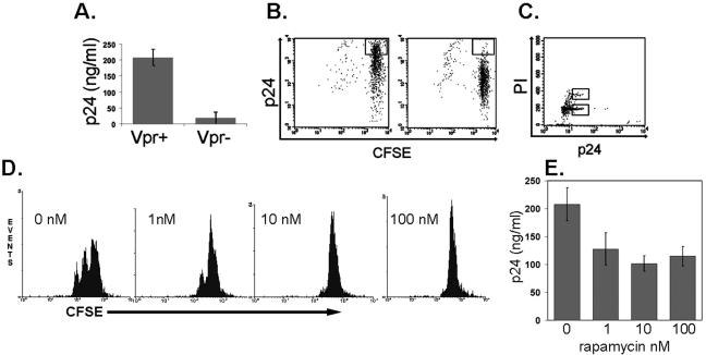
Characterization of the role of Vpr in enhanced HIV production. (A) Replication of wild-type versus Vpr− HIV in EC-T-cell cocultures 9 days postinfection as assessed by p24 measured in the supernatant by ELISA. (B) CFSE staining of infected cells harvested on day 9 postinfection. The box identifies the CFSEhigh p24high cells, which are fewer in cultures infected by Vpr− virus (right). (C) PI staining of infected cells harvested on day 9 postinfection. The lower box identifies cells in the G0/G1 state, and the upper box identifies the cells in the G2 state of the cell cycle. Note that the majority of cells are in G0/G1. (D) FACS analyses of CFSE-labeled CD4+ T cells treated with PHA-L and rapamycin. Note that the cells remain CFSEhigh. (E) Replication of wild-type HIV in EC-T-cell cocultures treated with rapamycin or vehicle (dimethyl sulfoxide, 0 nM rapamycin) 9 days postinfection as assessed by p24 measured in the supernatant by ELISA. The data in panels A to E are from one of three experiments with similar results. The error bars indicate standard deviations.

