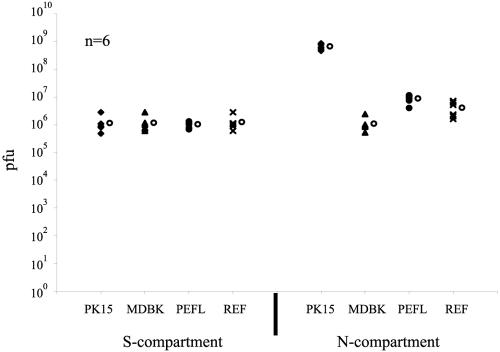
FIG. 4.
Neuron-to-cell spread of infection is not cell type specific. PK15 cells, Madin-Darby bovine kidney cells, pig embryonic fibroblasts, and rat embryonic fibroblasts were plated in the N-compartment. Neurons in the S-compartment were then infected with PRV Becker at high MOI for 48 h. Infected cells were then harvested from both the S- and N-compartments and titered on PK15 cells. Six chambers were used for each type of infection. The standard deviations are: N-compartment (PK15 cells, ±1.6 × 10⁸, MDBK cells, ±6.6 × 10⁵, pig embryonic fibroblasts, ±2.8 × 10⁶, rat embryonic fibroblasts, ±2.4 × 10⁶); S-compartment (PK15 cells, ±8.3 × 10⁵, MDBK cells, ±8.2 × 10⁵, pig embryonic fibroblasts, ±2.7 × 10⁵, rat embryonic fibroblasts, ±7.8 × 10⁵). The open circle beside each data set represents the average value for that particular set of data.

