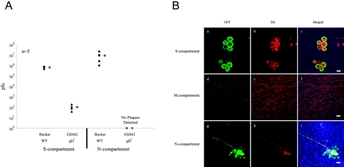
FIG. 6.
Neuron-to-cell transmission of infection does not require gD. A: Neurons in the S-compartment were infected at high MOI with either PRV Becker or GS442, a complemented gD null virus that expresses GFP. At 24 h postinfection, infected cells in the S- and N-compartments were harvested and titered in PK15 cells. Five chambers were used in each type of infection. The open circle beside each data set represents the average value for that particular set of data. The standard deviations are: N-compartment (Becker, ±1.0 × 10⁷); S-compartment (Becker, ±1.6 × 10⁵; GS442, ±5.9 × 101). B: Immunofluorescence experiment on PRV Becker- and GS442-infected neurons in the trichamber system. Culture samples in the S- (a to c), M- (d to f), and N-comparments (g to i) were labeled with antibodies against GFP (a, d, and g), DiI (b, e, and h), and the nuclear dye Hoechst shown in the merged image (c, f, and i). Scale bar: 20 μm.

