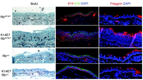
FIG. 1.
Proliferation and differentiation in RbWT/WT and RbΔ/Δ epidermis. Shown are immunohistochemistry and immunofluorescence images of RbWT/WT (top row), K14E7RbWT/WT (second row), RbΔ/Δ (third row), and K14E7RbΔ/Δ (bottom row) ear epidermis from 21-day-old mice. Left column: BrdU immunohistochemistry is brown with hematoxylin counterstain. Middle column: immunofluorescence stain for keratin 14 (K14, red) and keratin 10 (K10, green) with DAPI nuclear counterstain. Right column: immunofluorescence stain for filaggrin (red) with DAPI nuclear counterstain.

