
Figure 3.
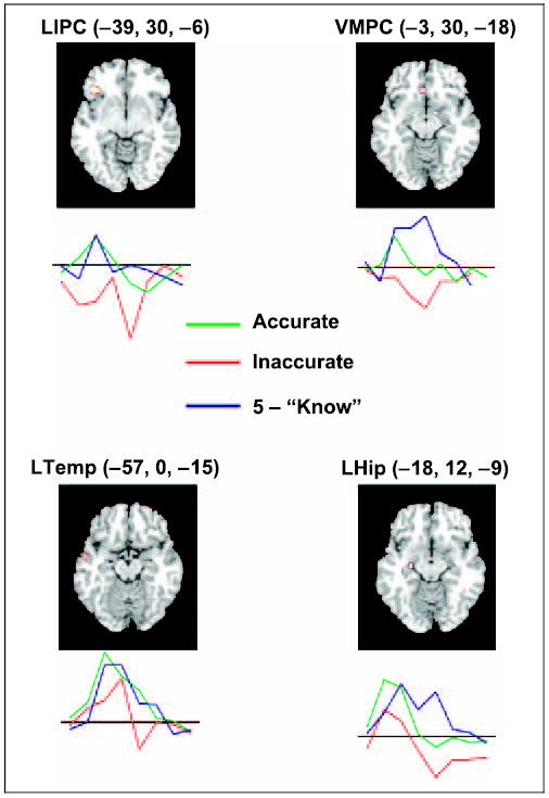
Four critical left hemisphere regions revealed by the contrast of accurate judgments > inaccurate judgments. The images and MNI coordinates indicate the local maximum. Below each region is a time-course graph of extracted signal levels used for SEM. The three conditions (accurate judgments, inaccurate judgments, and “know” trials) are indicated with separate colored lines and reflect data averaged across the identified cluster. The y-axis of this graph is normalized percent signal change and the x-axis is poststimulus time from 0 to 16 sec. All time courses are baselined to the average of −4 to 0 sec prestimulus onset. LIPC = left inferior prefrontal cortex; LHip = left hippocampus; LTemp = left temporal cortex; VMPC = ventral medial prefrontal cortex.