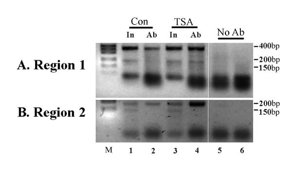
Figure 2.
Chromatin immunoprecipitation assay of histone acetylation at the H19 locus in treated rhabdomyosarcoma cells. Cells were either treated (TSA) or not treated (Con; control) with histone deacetylase inhibitor (500 nM TSA for 6 hrs). DNA and proteins were cross-linked and precipitated with an antibody to acetylated histone 4, prior to reversing cross-links, amplifying DNA with specific primers and separating the products on a gel. (A) Results obtained using primers specific for the transcribed region of the H19 gene (Region 1), which give an expected band size of 355 bp. Equal amounts of input DNA (In) were used in each immunoprecipitation (lanes 1 and 3) and a no antibody control was run for each sample (lanes 5 and 6). Treatment with TSA gives an increase in signal for the 355 bp target, indicating an increase in acetylated histones associated with the transcribed region (lanes 2 and 4). (B) Primers for a region in the insulator (Region 2) upstream of H19 and downstream of IGF2 give a specific band of 161 bp and this also shows a marked increase after TSA treatment of the cells. Controls are as for (A) above. M; 1 kb DNA size marker (Life Tech.). Minor bands present in some lanes represent non-specific artifacts: primers are visible at the bottom of the gel. Negative images of ethidium-stained gels are shown.

