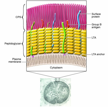
Figure 1.
Cell wall structure of GBS. Electron micrograph image of a type III GBS organism labeled with antiserum raised to whole organism; the inset shows detailed representation of important surface structures. The GBS cytoplasm is bounded by an inner cell membrane. Surrounding this membrane is a peptidoglycan layer that anchors the negatively-charged CPS and group B antigen polysaccharide. Extending from the cell membrane are lipoproteins and glycolipids, including an anchor for LTA; LTA is a structure composed of a repeating carbohydrate phosphate polymer. Most surface proteins attach to the peptidoglycan. CPS, LTA, and cell-surface proteins contribute to the organism's ability to adhere to and invade host cells and evade host immune defenses in the course of pathogenesis. Data reported by Doran et al. (15) in this issue of the JCI reveal that an enzyme involved in the synthesis of a glycolipid anchor is required for normal GBS invasion of host cells in vitro and for GBS virulence in vivo.

