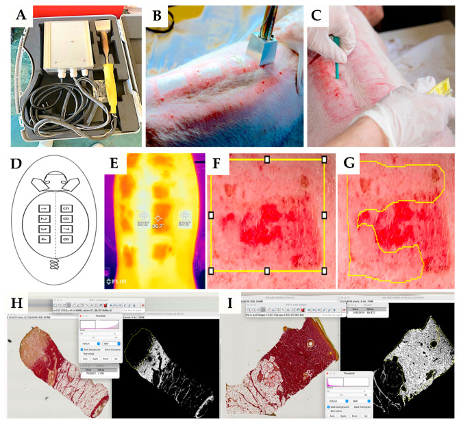
Figure 6.
Illustrations of the experimental procedure. Heater and its application (A,B); technique of tissue sampling (C); schema of burn place application (D); representative photograph of skin and wound using an infrared camera (E); measurement of the whole area (yellow borders on picture (F)) and reepithelialization area (yellow borders on picture—(G)) of the wound on 30th day post-thermal injury; measurement and quantification of collagen density in wound tissue using Picrosirius red staining (H,I).

