Fig. 3.
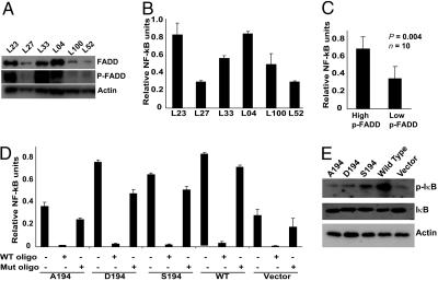
Induction of NF-κB activity by p-FADD. (A) Lysates from lung tumor samples expressing high (L23, L33, and L04) and low (L27, L100, and L52) p-FADD were subjected to Western blot analysis using anti-FADD, anti-p-FADD, and anti-actin antibodies. (B) Lysates from nuclei-enriched fractions were prepared from high (L23, L33, and L04) and low (L27, L100, and L52) p-FADD-expressing tumors samples, and NF-κB activity was monitored by using the functional NF-κB TransAM kit from Active Motif. Data shown is derived from mean (±SEM) of relative NF-κB of three separate experiments performed in triplicate. (C) NF-κB activity was significantly higher in tissues containing high levels of p-FADD, as compared with those having low levels of p-FADD (data derived from A and B; n = 10, P = 0.004). (D) Nuclear lysates from various Jurkat cell lines expressing FADD or mutant FADDs were prepared, and NF-κB activity was monitored. For competition experiments, lysates were preincubated with either WT NF-κB consensus oligonucleotide or mutant NF-κB oligonucleotide, and NF-κB activity was then assessed. (E) Western blot analysis of whole-cell lysates obtained from the WT FADD or various mutant FADD-expressing Jurkat cell lines by using anti-I-κB, anti-phospho I-κB, and anti-actin antibodies.

