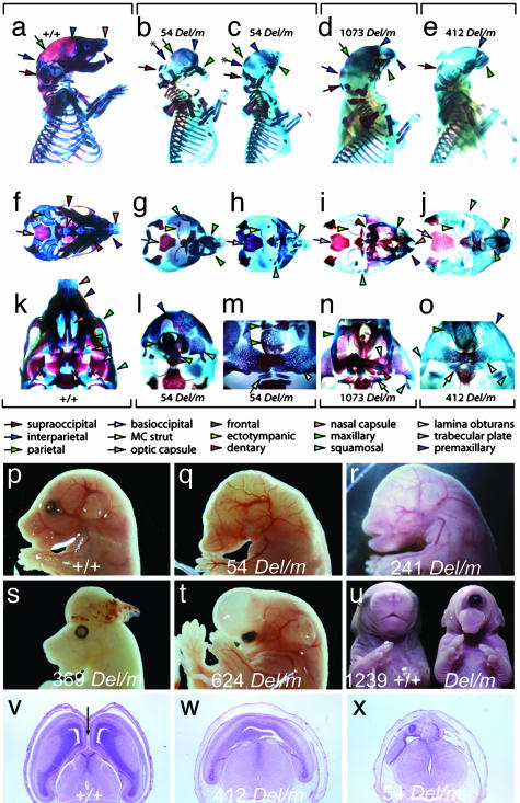
Fig. 2.
Craniofacial and forebrain development. The genotype of mutant animals is represented as Del/m to indicate the mutation (m) in trans to the Del(13)Svea36H chromosome (Del). (a-o) Differential bone and cartilage staining of wild type (+/+; a, f, and k) and mutants from three lines: 54 Del/m (b, c, g, h, l, and m), 1073 Del/m (d, i, and n), and 412 Del/m (e, j, and o). Norma lateralis views (a-e) demonstrate that each mutant specimen is microcephalic and has significant neurocranial, splanchnocranial, and dermatocranial defects, whereas norma basalis views (f-o) highlight the nature of the midline defects of the mutants. The nasal and optic capsules are severely deficient, as are the trabecular basal plate of the neurocranium to which they normally attach and the dermatocranial elements that develop in association with the capsules. Labeled colored arrows represent keys to the identification of homologous elements. (p) Lateral view of the cranial region of a wild-type 15.5-dpc embryo. (q) Lateral view of the cranial region of a line 54 15.5-dpc Del/m embryo showing impaired craniofacial development with anophthalmia, agnathia, and absence of ear pinnae. (r) Lateral view of the cranial region of a line 241 15.5-dpc Del/m embryo showing anophthalmia and absence of ear pinnae. (s) Lateral view of the cranial region of a line 369 15.5-dpc Del/m embryo exhibiting exencephaly. (t) Lateral view of the cranial region of a line 624 15.5 dpc Del/m embryo exhibiting a single, ventrally displaced eye beneath a thin walled holosphere. (u) Anterior view of an 18.5-dpc wild-type embryo (Right) and a 1239 Del/m embryo with cyclopia (Left). (v-x) Transverse sections through the prosencephalon of a 15.5-dpc wild-type embryo (v) or hemizygous mutants (w and x) showing hypoplastic lateral ventricles in lines 412 and 54 with no development of the interhemispheric fissure (arrow in v).

