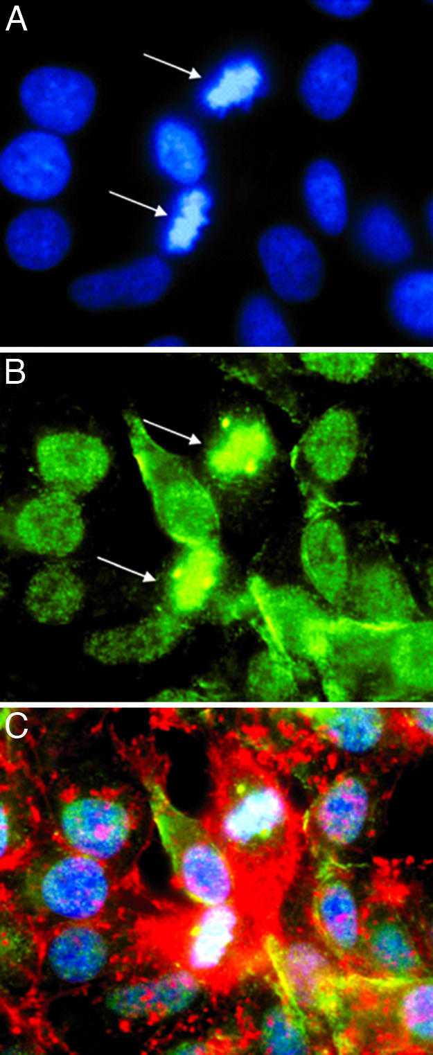
Fig. 4.

Localization of β-adducin in centrioles of PTN-treated dividing HeLa cells. (A) PTN-treated (50 ng/ml) HeLa cells stained with DAPI (blue). (B) PTN-treated (50 ng/ml) HeLa cells stained with anti-phosphoserine 713 and 726 β-adducin antibodies (green). (C) PTN-treated (50 ng/ml) HeLa cells stained with phalloidin (red), anti-phosphoserine 713 and 726 β-adducin antibodies (green), and DAPI (blue). Note the localization of phosphoserine 713 and 726 β-adducin to chromatin and the centrioles during metaphase (arrows).
