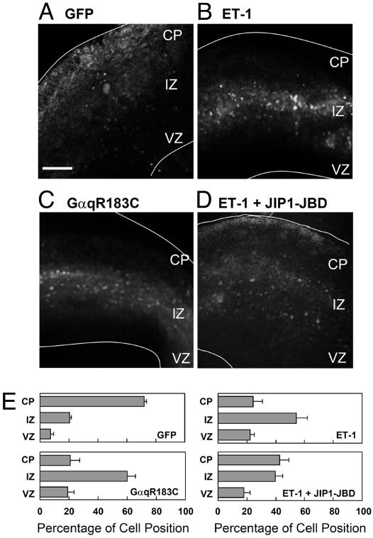
Fig. 8.
Effect of GPCR signaling on the migration of GFP-labeled cells originating from the VZ in a slice culture. Cortical slices were prepared from E16.5 mouse telencephalon. (A–D) Cells in the VZ were infected with adenoviruses harboring GFP alone (A and B), GFP plus GαqR183C (C), or GFP plus JIP1-JBD (D). Slices were cultured for 3 days in vitro without (A and C) or with 1 μM ET-1 (B and D). The white lines represent pial and ventricular surfaces. (E) Quantitative analysis of radial migration in A–D is shown. The slice was subdivided into three regions, indicated as CP, IZ, and VZ. Each score represents the mean percentage of relative intensity ± SD; n = 5. (Bar, 250 μm.)

