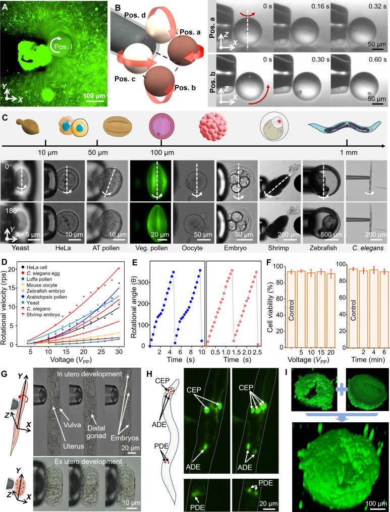
Fig. 4. Multidirectional rotations in 3D axisymmetric streaming and related applications.
(A) Streaming around the trapped microspheres visualized by the 1-μm PS fluorescent particles (movie S4). Pos., position. (B) Multidirectional rotation by altering the object’s relative position in the axisymmetric vortex (movie S4). The microsphere at positions a and b rotates along the Z axis and the Y axis, respectively. (C) The μSonic-hand traps and rotates yeast cells (~7 μm), HeLa cells (~20 μm), A. thaliana pollen (~30 μm), luffa pollen (~60 μm), mouse oocytes (~100 μm), mouse embryos (eight-cell stage, ~120 μm), shrimp eggs (~250 μm), zebrafish embryos (~1 mm), and C. elegans (~50 μm in diameter and 1 mm in length) (movie S5). AT, A. thaliana; Veg., luffa (vegetable) pollen. (D) Rotational velocity versus input voltage. (E) Rotation angle of mouse embryos and A. thaliana pollen over time. (F) Results of cell viability after μSonic-hand manipulation at different driving voltages and operating times. (G) 3D observation of C. elegans development by rotating their embryos from in utero to ex utero (movie S5). (H) Robust immobilization and controllable rotation of C. elegans (BZ555) allow direct observation of the dopaminergic neurons of interest from different directions (movie S5). CEP, cephalic; ADE, anterior deirid; PDE, posterior deirid. (I) Enhanced 3D images of a cell spheroid by integrating two images captured from 0° and 180° using a standard laser scanning confocal microscopy (LSCM) (movie S5).

