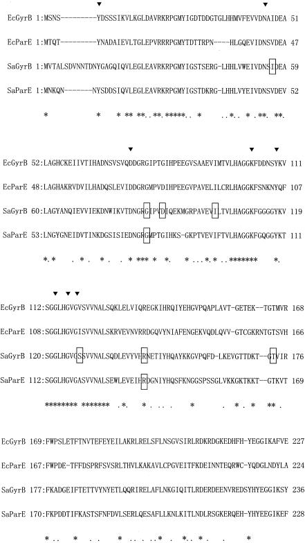
FIG. 2.
Alignment of GyrB and ParE N-terminal amino acid sequences. Triangles, residues from E. coli GyrB region involved in ATP binding (26); asterisks and dots under the amino acid sequences, identical and similar residues in all four proteins, respectively; dashes, gaps introduced to maximize similarities; boxes, amino acid sequences that underwent changes in the novobiocin-resistant mutants; Ec, E. coli; Sa, S. aureus.

