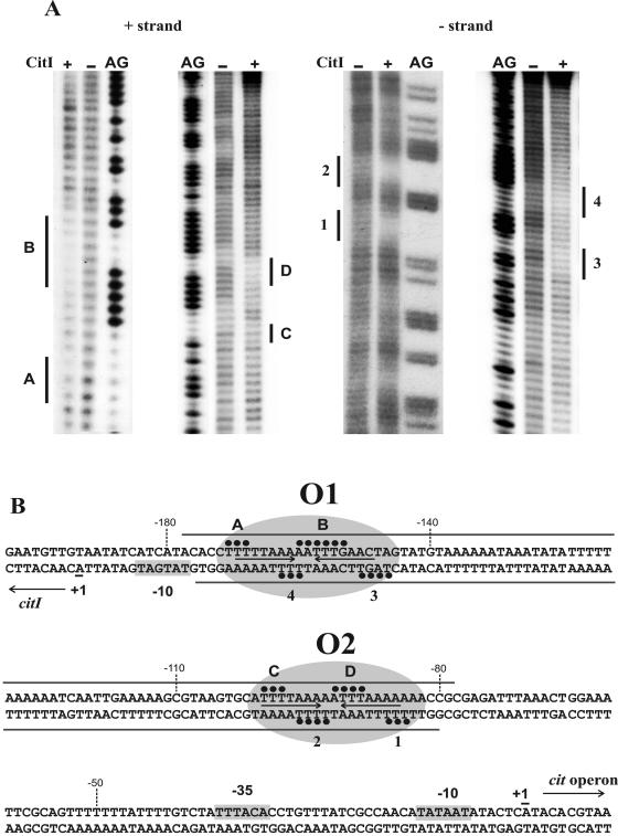
FIG. 2.
Protection of the DNA sequence containing PcitI and Pcit promoters by CitI. (A) Hydroxyl radical footprinting of CitI binding. A 150-bp DNA probe including the DNase I protected region (see Fig. S1 in the supplemental material) was γ-32P labeled in either the plus or the minus strand (see details in Materials and Methods). The assays were performed in the presence (+) or absence (−) of CitI. The footprintings of the plus (left panel) and minus (right panel) strands are depicted, and they correspond to CitI-DNA complex II. Regions protected from the cleaving agent by CitI on the plus strand (A, B, C, and D) and on the minus strand (1 to 4) are indicated. Footprintings are flanked by the corresponding G+A sequence ladders (AG) generated with the same DNA probe (see details in Materials and Methods). (B) Schematic representation of DNA regions protected by CitI. Closed circles and gray bars indicate protections by CitI obtained, respectively, in the hydroxyl radical (A) and DNase I (see Fig. S1 in the supplemental material) footprintings. The inverted repeats identified in the CitI binding sites are shown with arrows. The operator sites identified, named O1 and O2, are enclosed in ovals. The transcriptional start sites (+1) identified in W. paramesenteroides (14) and the putative −10 and −35 boxes of the Pcit and PcitI promoters are depicted.

