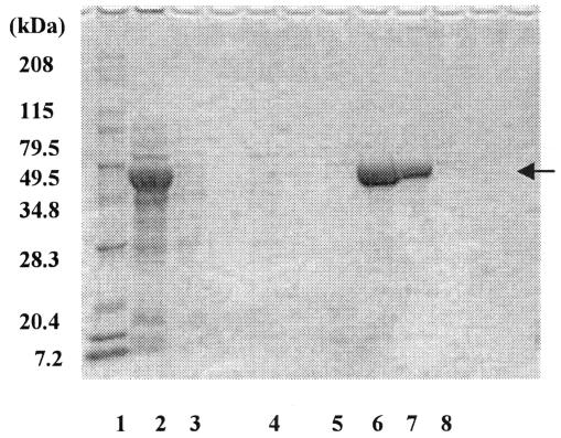
FIG. 2.
SDS-polyacrylamide gel electrophoresis analysis of cell lysates and purified enzyme fractions. Lane 1, marker; lane 2, crude cell extracts; lanes 3, 4, and 5, fractions eluted from the nickel column with an imidazole buffer of 10 mM, 40 mM, and 100 mM, respectively; lanes 6 to 8, fractions eluted with 500 mM imidazole buffer (amounts of protein applied to the SDS gel were different).

