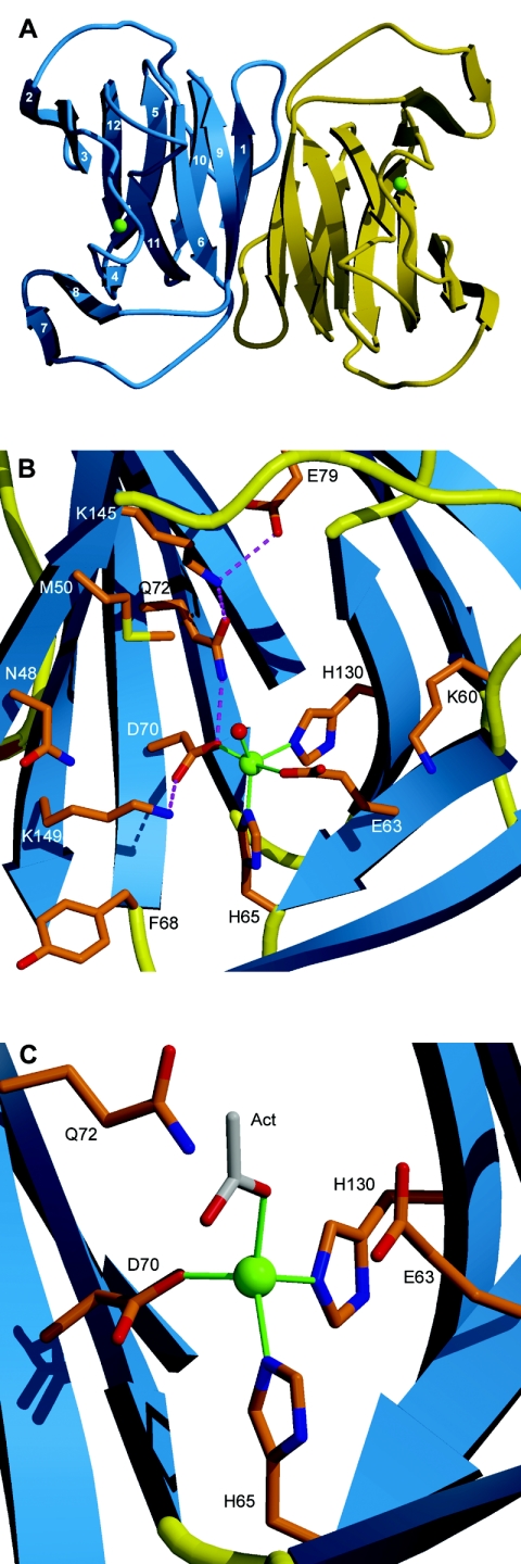
FIG. 1.
Crystal structure of YhcH. (A) Ribbon diagram of the dimeric molecule of YhcH. β-Strands are represented by arrows and are numbered consecutively. Cu ions are indicated by green spheres. (B) Putative active site in subunit B of YhcH as viewed from the left with respect to panel A. (C) Cu coordination in subunit A. The acetate molecule is gray. Amino acids are labeled with their one-letter abbreviations. Cu coordination bonds are indicated by green lines, and the hydrogen bond network is indicated by dashed magenta lines.

