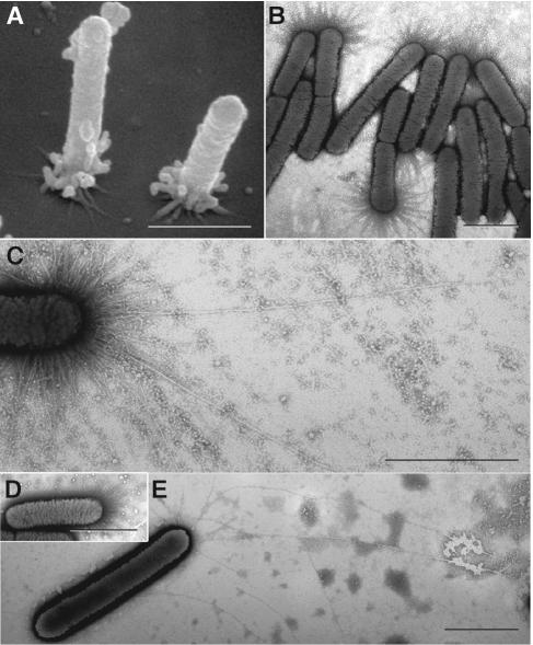
FIG. 4.
Pili of wild-type and mutant X. fastidiosa. (A) Scanning electron micrograph of wild-type cells attached to the substratum at the pilus-bearing polar ends. Many pili degenerated into globular masses during specimen preparation. (B and C) Transmission electron micrographs of negatively stained (phosphotungstic acid) wild-type cells depicting an abundance of short pili (B), and fewer long type IV pili (C). (D and E) Negatively stained preparations of mutants 1A2 and 6E11 depicting only short pili and only longer (type IV) pili, respectively. Pilus lengths are noted in Fig. 8. Bars, 1 μm.

