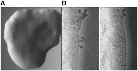
FIG. 5.
Xylella fastidiosa mutant characteristics. (A) Light micrograph of X. fastidiosa mutant 1A2 colony on agar medium with a well-demarcated fringeless periphery. (B) Sequential images (0 min and 20 h 19 min, respectively) depicting progressive expansion of the 1A2 colony (see movie 1B in the supplemental material; also see expanded versions and additional movies at http://www.nysaes.cornell.edu/pp/faculty/hoch/movies/). Bar, 20 μm.

