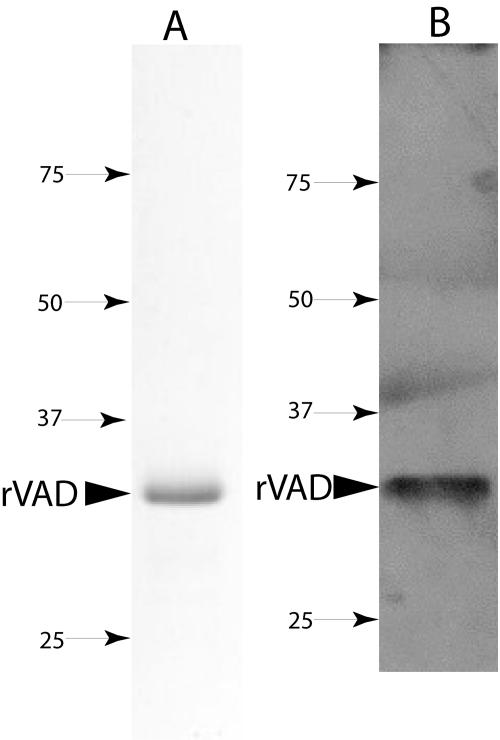
FIG. 1.
SDS-PAGE of the purified SpoVAD fusion protein (A) and detection of the SpoVAD fusion protein with anti-His tag antibody (B). (A) The SpoVAD fusion protein was purified from the pellet fraction of induced E. coli as described in Materials and Methods. An aliquot (∼10 μg) of the purified protein was run on SDS-PAGE and the protein visualized by Coomassie blue staining. (B) Approximately 0.5 μg of the SpoVAD fusion protein was run on SDS-PAGE, transferred to a polyvinylidene difluoride membrane, and detected with anti-His tag antibody as described in Materials and Methods. In panels A and B, the arrowhead denotes the purified SpoVAD fusion protein (∼32 kDa) and numbered arrows show the migration positions of molecular mass markers (sizes are in kilodaltons). Note that the apparent bands in panel B above the 37- and 50-kDa markers do not actually fall in the lane where the sample was run and are only smudges on the X-ray film.

